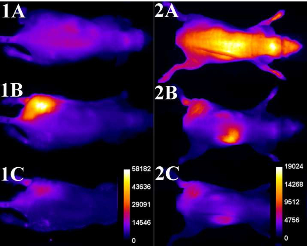
Figure 4.
Typical infection imaging montages. Nude, athymic mice were injected with S. aureus (~108 CFU) in LB broth (50 µL) in the left leg and sterile LB broth (50 µL) in the right leg. Six hours later, 10 nmol of probe 1 (series 1) or probe 2 (series 2) was injected via the tail vein, and whole-animal dorsal images were acquired at: [A] 0, [B] 3, and [C] 12 hours after probe dosage. The intensity scale bars (a.u.) apply to all images in each series.

