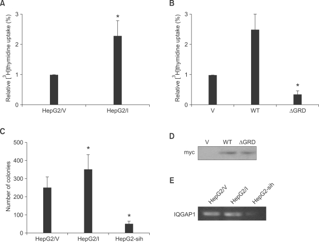
Figure 2.
Effect of IQGAP1 on proliferation of HepG2 cells. (A) HepG2 cells (2 × 10⁵) stably expressing control vector pcDNA3 (HepG2/V) or Myc-tagged IQGAP1 (HepG2/I), were seeded into 24-well culture dishes. After 24 h, [3H]thymidine was added for 18 h, and incorporation measured. All assays were performed in triplicate. The means ± S.D. are shown (n = 12 replicates/group). *P < 0.01. (B) Equal numbers of HepG2 cells were transiently transfected with 10 µg of vector (V), wild type (WT) IQGAP1, or IQGAP1ΔGRD (ΔGRD). Cell proliferation was quantified as described for A. Data are expressed as means ± S.D. [3H]thymidine uptake relative to V cells, (n = 3, in quadruplicate). *P < 0.01; **P < 0.05. (C) HepG2/V, HepG2/I, and HepG2-sih (1 × 10⁴) cells were assessed by soft agar assay after 14 days at 37℃. Colonies > 0.2 mm were counted using a light microscope. The data are expressed as means ± S.D, n = 2, performed in triplicate. *P < 0.01 relative to HepG2/V-derived colonies. (D) Expression level of transfected IQGAP1 in HepG2 cells that transfected with 10 µg of vector (V), wild type (WT) IQGAP1, or IQGAP1ΔGRD (ΔGRD) was detected by Western blot using myc antibody. (E) The mRNA level of IQGAP1 in HepG2/V, HepG2/I, and HepG2-sih (1 × 10⁴) cells was accessed by RT-PCR.

