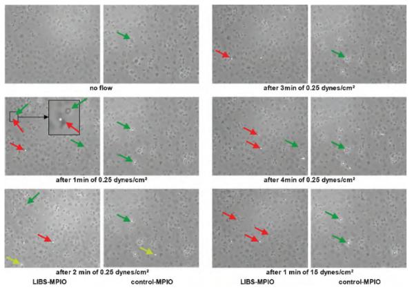
Figure 4.
Flow chamber experiments under venous flow conditions (0.25 dyn/cm2) and strong arterial flow conditions (15 dyn/cm2). Microscopic view of iron oxide microparticle (MPIO) binding for six different time points (no flow; 0.25 dyn/cm2 after 1 minute, 2 minutes, 3 minutes, and 4 minutes; and 1 minute of 15 dyn/cm2). The inset shows a zoomed area of interest with a neighboring MPIO (red arrow) and a platelet (green arrow). The MPIO appears as a white and round structure, which is also attached on the surface of a flattened and therefore activated platelet (dark structure underneath). Conversely, the platelet marked with the green arrow is not flattened but has a round morphology, which is slightly bigger than the MPIO and has a dark halo. This corresponds to a platelet, which is not flattened but only partially attached to the well plate and is therefore not yet completely activated; the number of such nonattached structures increases with higher flow and shear stress. The yellow arrow indicates a flowing MPIO. Ligand-induced binding site MPIO binding increases over time and remains constant after switching from venous to arterial flow conditions, whereas only modest background binding of control MPIOs to activated platelets was observed.

