
Fig. 9.
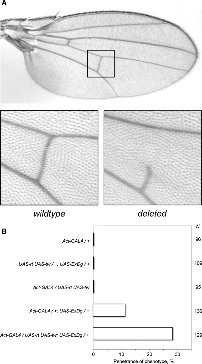
Effect of ExDG ectopic expression on the posterior crossvein development. (A) Top panel, wild-type Drosophila wing with the region of posterior crossvein shown by a rectangle. Bottom panels show enlarged examples of a wild-type and a defective crossvein (with deleted anterior part) as a result of ExDG ectopic expression using an Act-GAL4 driver. The phenotype varied from a severe ‘deletion’ of the crossvein (shown) to a less extreme but always obvious loss of crossvein tissue. (B) Penetrance of the crossvein defect is enhanced by RT-TW activity. N indicates the number of flies scored for each genotype. ExDG encompasses the complete extracellular domain of DG-C (a.a. 1–1048).