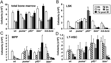
Figure 3.
Loss of Puma prevents stem cell apoptosis upon IR. (A) Cellularity of both femora and tibiae was assessed in mice of the indicated genotypes after exposure to a single dose of IR. (B–D) Cell counting and staining with cell surface marker-specific antibodies were used to identify and enumerate the different stem cell populations. Bars represent mean ± SEM of three to six animals per genotype and time point from three independent experiments. (*) LSK cell numbers were significantly different between wild-type or bim−/− versus puma−/− (P < 0.0002) or p53−/− mice (P < 0.0014) at all time points analyzed after IR; MPP numbers were different between wild-type or bim−/− versus puma−/− (P < 0.04) or p53−/− mice (P < 0.035) at days 1, 3, and 7 after IR; LT-HSC numbers were different between wild-type or bim−/− versus puma−/− (P < 0.038) or p53−/− mice (P < 0.003) at day 7 after IR. (§) In untreated mice, LT-HSC numbers were different between bim−/− and all other genotypes (P < 0.05).

