Figure 5.
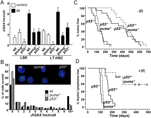
Replication stress-associated DNA damage in wild-type HSCs after IR and restoration of thymic lymphoma development in puma−/− mice by concomitant loss of p53. (A) Quantification of DNA damage foci in LSK cells and LT-HSCs derived from mice of the indicated genotypes. Six weeks after the fourth cycle of IR, stem cells were sorted from the bone marrow onto poly-L-lysine-coated coverslips and stained for γH2AX. (B) Quantification of the percentage of LT-HSCs containing a given number of γH2AX foci. (Insert) Immunofluorescence detection of γH2AX foci in LT-HSCs and LSK cells derived from wild-type, puma−/−, and p53−/− mice 6 wk after the last dose of irradiation. (Green) γH2AX; (blue) DAPI. Images were acquired with a Leica confocal scanning microscope, and the numbers of foci per cell were quantified by using the CellProfiler software, measuring an average of 100 cells per sample from four to six mice per genotype. (*) IR wild type versus puma−/− or p53−/− (P < 0.001). LT-HSCs from untreated p53−/− mice show reduced foci number (P < 0.05). (B) Assessment of foci number per cell, in relation to the total number of cells analyzed. Cohorts of p53+/− and p53−/− mice proficient or deficient for puma were monitored for spontaneous tumorigenesis and tumor formation triggered by fractionated IR. (C,D) Kaplan-Meier analysis of tumor-free survival of untreated p53+/− (n = 15), p53−/− (n = 15), p53+/−puma−/− (n = 16), and p53−/−puma−/− (n = 15) mice (C), or thymic lymphoma-free survival of irradiated p53+/− (n = 10), p53−/− (n = 9), p53+/−puma−/− (n = 9), and p53−/−puma−/− (n = 5) mice (D). Mean survival in days ± SE: 176 ± 8 for p53+/− versus 307 ± 37 for p53+/−puma−/−; P < 0.0001.

