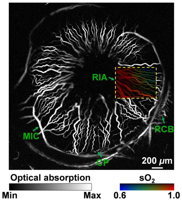
Figure 3.

Label-free photoacoustic ophthalmic angiography of hemoglobin oxygen saturation (sO2) in the iris microvasculature of a living adult Swiss Webster mouse. The imaged region is the same as that shown in Fig. 2a, but with a focal plane closer to the posterior segment to better visualize the peripheral vascular structures. A dual-wavelength (570 and 578 nm) sO2 measurement was performed on the boxed region of interest. A vessel-by-vessel sO2 mapping was generated and overlaid on the maximum amplitude projection image acquired at 570 nm. CP: ciliary process; MIC: major iris circle; RCB: recurrent choroidal branch; RIA: radial iris artery.
