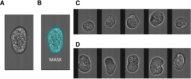
FIG 1 .
Measurement of amebic morphology. The Amnis Imagestream imaging cytometer was used to measure the morphology of fixed amebic trophozoites. (A) Bright-field image of an elongated trophozoite. (B) The pixels which constituted the bright-field image of the trophozoites (MASK). From the mask, the computational features of area and circular morphology were derived. A high circularity score resulted from an internally consistent measurement of cell radius. Representative images show trophozoites characterized and “tagged” as either circular (C) or elongated (D).

