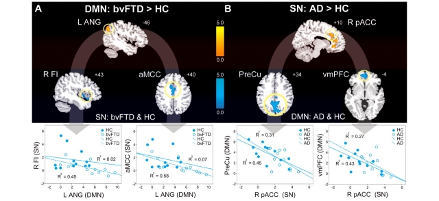
Figure 4.
ICN enhancements relate to specific connectivity disruptions within the reciprocal networks. (A) Left angular gyrus (L ANG) DMN connectivity was intensified in patients with bvFTD versus healthy controls and the 3 mm radius spherical region of interest centred on its peak showed a significant inverse correlation with SN connectivity in right frontoinsula (R FI), anterior mid-cingulate cortex (aMCC) and the dorsal pons (not shown) across patients with bvFTD and healthy controls (HC). Group-wise scatterplots illustrate the relative contribution of each group to these relationships for each anticorrelated region. (B) Right pregenual anterior cingulate cortex (R pACC) SN connectivity, enhanced in patients with Alzheimer’s disease (AD), showed a significant inverse relationship with DMN connectivity in precuneus (PreCu), ventromedial prefrontal cortex (vmPFC) and bilateral middle temporal gyrus (not shown) across patients with Alzheimer’s disease and healthy controls. Results are atrophy-corrected and thresholded at a joint height and extent probability of P < 0.05, corrected at the whole brain level. Colour bars represent t-scores, and statistical maps are superimposed on the Montreal Neurological Institute template brain.

