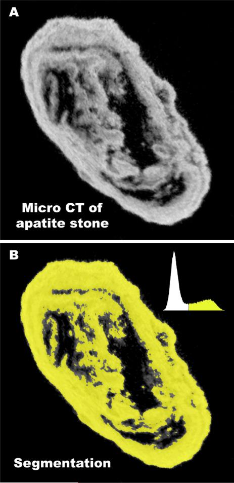
Fig. 1.
Method for determining volume of x-ray lucent regions (voids) using micro CT. A: Micro CT slice of an apatite stone, showing dark regions (voids) within the stone. Total micro CT reconstruction of this stone involved a stack of 243 such slices, each representing 25.5 μmof thickness through the stone. B: Stone mineral was segmented using image histogram, shown in upper right of panel. Segmented regions were summed in all slices to give total stone mineral volume. This sum was repeated, but including void regions to give total stone volume, and void volume calculated from the difference.

