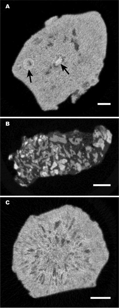
Fig. 3.
Micro CT slices of three ‘odd’ morphologies for apatite stones, found among the 15 stones in this study. A: Stone was relatively solid, with isolated nodule-like structures within it (arrows). B: Stone was composed of small pieces of apatite, each <0.5 mm,held together with an x-ray lucent material. C: Stone contained many small voids, which were arranged radially about the stone axis. By FT-IR, this stone showed the presence of some amorphous calcium phosphate. Bars indicate 1 mm.

