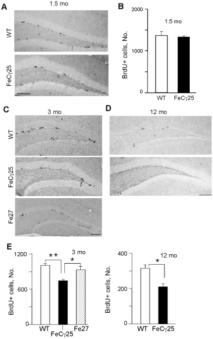
Figure 1. AICD transgenic mice show decreased cell proliferation in the SGZ of hippocampus.
Representative images of BrdU immunostaining in the subgranular zone (SGZ) of the dentate gyrus performed on 1.5-mont-old (A) 3-month-old (C) and 12-month old (D) wild-type and FeCγ25 animals one day after the final BrdU injection (100 mg/kg). (A–B) At 1.5 mo there was no statistically significant difference in the number of BrdU-labeled cells between wild-type and FeCγ25 mice, quantified in (B). (E) Quantitative analysis of the total number of BrdU+ cells throughout the entire rostro-caudal extent of the hippocampus in 3-month-old (top) and 12-month-old (bottom) animals reveal a statistically significant decrease in the number of BrdU+ cells in the SGZ of FeCγ25 mice compared to wild-type mice. (n = 3, 4 and 4 for both wild-type and FeCγ25 mice at ages 1.5 mo, 3 mo and 12 mo respectively. n = 3 for Fe27 mice at 3 mo. * p<0.05, ** p<0.01).

