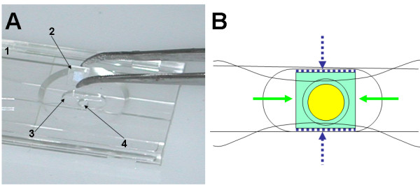
Figure 3.

Attachment of the glass picowells substrate to the i3C base. (A) The base (1) is placed upside down, and the picowells substrate (2) is gently placed in its socket (3, niche) with its picowells facing down the lower entrance of the cell chamber (4). Next (B): Medical grade UV adhesive (dashed blue) is gently injected in the empty space created between the chip edge and the niche wall, then irradiated and cured. Then, a small drop of liquid PDMS (green) is gently poured near the free edges, and is drawn by capillary forces to fill the empty space between the peripheral face of the chip and the bottom of the niche. The PDMS does not penetrate into the cell chamber area (yellow) due to lack of capillary forces in this open region. When PDMS is cured, the structure is practically sealed to fluids and withstands freezing - thawing cycles.
