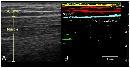
Figure 3. Delineation of Tissue Layers and Zones.
(3A.) Ultrasound image. This longitudinal image of the thigh shows the subcutaneous and muscle layers. Echogenic bands (white bands) are seen in the subcutaneous layer. (3B.) Thresholded image. Thresholding the image delineates the echogenic bands in the dermal, SQ, and perimuscular zones.

