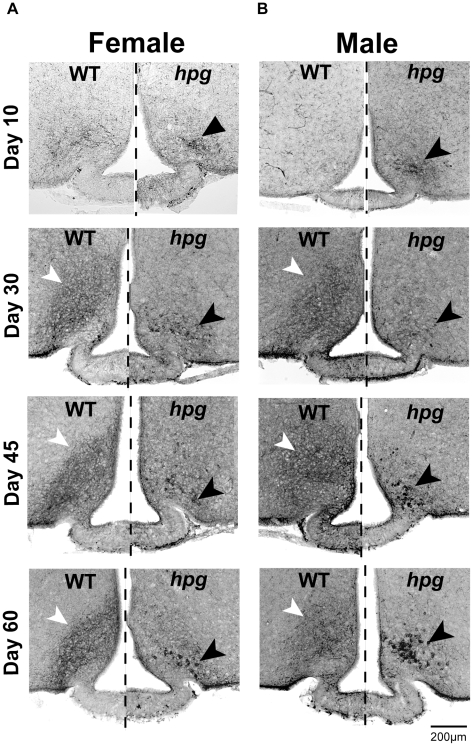
Figure 4. Altered distribution pattern of kisspeptin immunostaining in the ARC of hpg mice.
Kisspeptin ICC across postnatal development (age 10, 30, 45 and 60 days) in WT and hpg females (A) and males (B). Kisspeptin ICC staining pattern in the ARC of both female and male WT mice (left of dashed line) is dominated by densely stained fibers that obscure kisspeptin-positive cell bodies (white arrowheads). In hpg mice (right of dashed line), ARC kisspeptin ICC shows reduced fiber staining and increased clusters of large, darkly stained cell bodies (black arrowheads). Scale bar = 200 µm.

