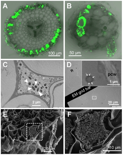
Figure 2. Root transverse sections and electron micrographs of tomato and Arabidopsis show GFP E. coli in the apoplast and inside root cells.
E. coli was detected inside tomato roots (A, C and D, E and F) and Arabidopsis roots (B). (A and B) Fluorescent images of transverse sectioned roots taken by CLSM. (C and D) Images taken by a transmission electron microscope. White triangles in (C) indicate E. coli cell present in apoplast. (D) Roots were probed with immunogold-labeled anti-GFP revealing E. coli in root cortex cells. Sub-image in (D) is a detail of dash-white square box. Gold labeling is marked with white arrows. Rhizodermis cell (R) and plant cell wall (pcw) is indicated. (F) is a detail image of (E) showing plant cells containing E. coli, and both images were taken by SEM.

