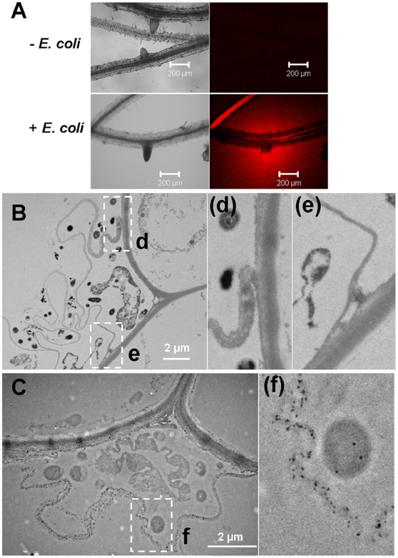
Figure 4. Root produced cellulase and extended the cell wall when incubated with E. coli Bl21.
(A) Incubation of Arabidopsis roots in 31 µg/mL resorufin cellubioside after incubating overnight with E. coli. After 2 h incubation, roots were viewed by CLSM. (B) TEM image of cell wall-like structure of plant roots encompassing bacteria. (C) TEM image of cellulase-gold labeling on the root sections with double labeling with the anti-GFP antibody. The size of the gold particle on bacteria is 15 nm (Au-particle specific to GFP E. coli) and gold particles on the plant material are 10 nm (Au-particle specific to plant cellulose). (d), (e) and (f) are detail images of insets d, e and f.

