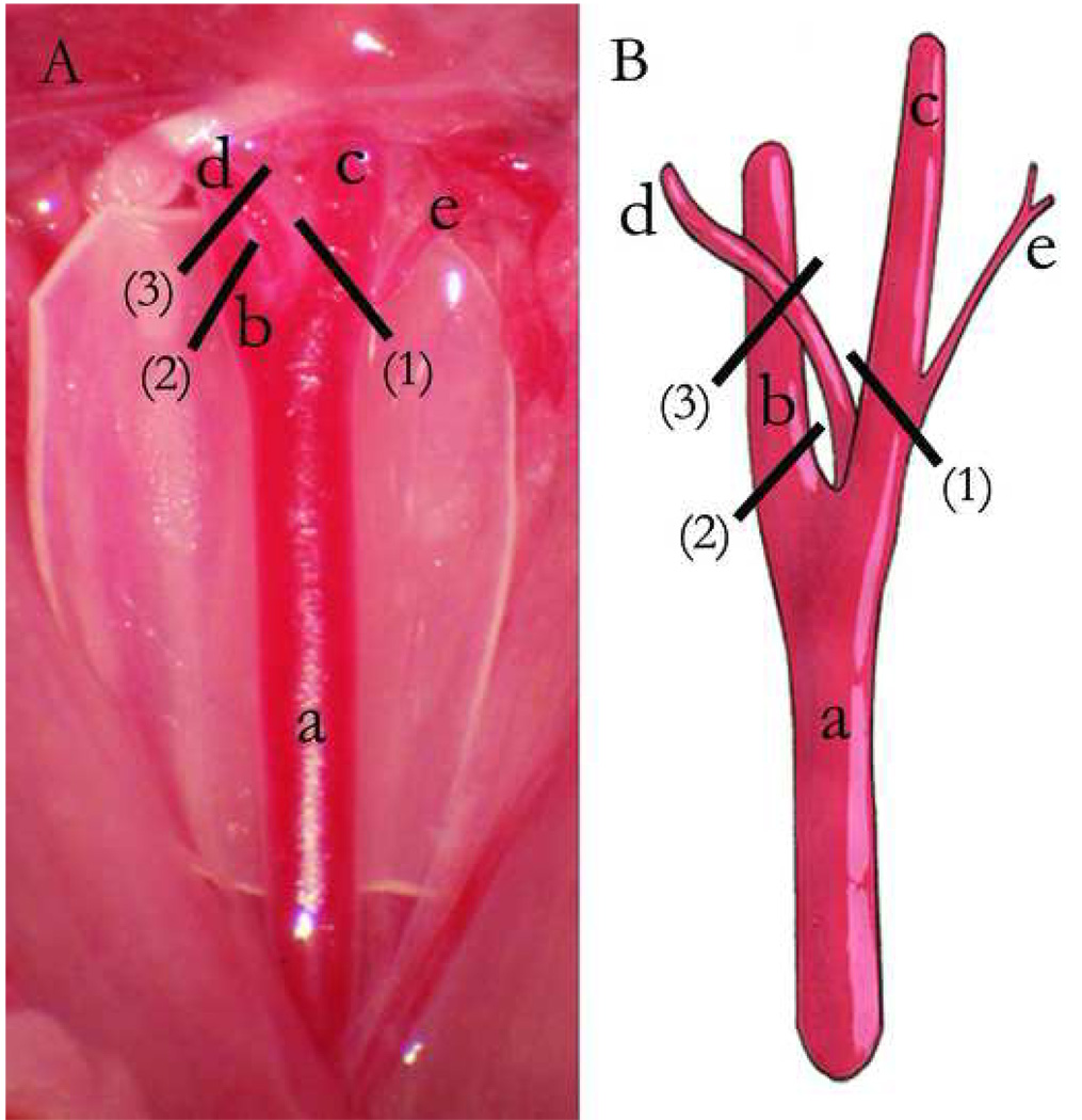
Fig 2.
Mouse carotid artery anatomy and partial ligation methods (A, Photographed right carotid anatomy (cut surgical glove background). B, Schematic representation). a, Right common carotid artery. b, Internal carotid artery. c, External carotid artery. d, Occipital artery. e, Superior thyroid artery. (1), Ligation of external carotid artery, including superior thyroid artery, but not occipital artery. (2), Ligation of internal carotid artery. (3), Ligation of internal carotid artery plus occipital artery.

