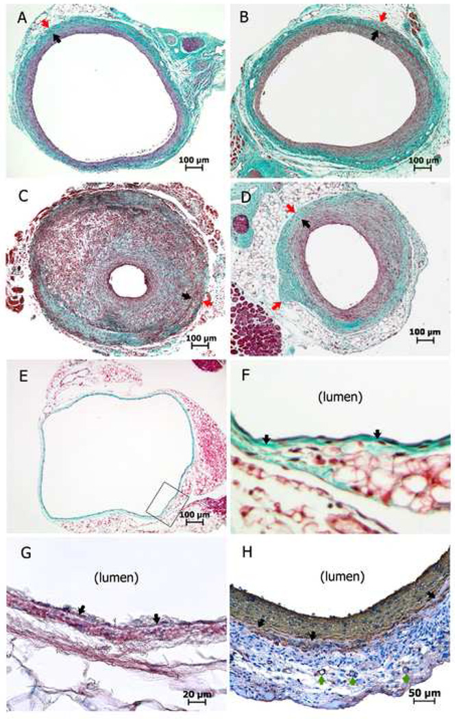
Fig 3.
Masson trichrome staining (A–F) and α-actin immunohistochemistry staining (G, H) of cross-sections of mouse vein grafts/vein. Black arrows depict the internal elastic lamina (A–D, F–H); red arrows show the tunica adventitia/peri-vascular tissue boundary (A–D); and green arrows show the vasa vasorum (H). A, Normal flow vein graft at day 28. B, Vein graft with distal arterial partial (internal carotid + occipital artery) ligation at day 28. C, Vein graft with distal arterial partial (external carotid + internal carotid) ligation (leaving the occipital artery patent) at day 14. D, Negative wall remodeling in a mouse vein graft at day 28. E, Normal IVC. F, Enlarged area defined by the black box of Fig 3E. G (normal IVC) and H (mouse vein graft at day 28), note some rose-red or brownish-red positive expression of α-actin in the tunica media (beyond the internal elastic lamina).

