
Figure 6. Survival studies of athymic nude mice implanted with human U87ΔEGFRvIII xenografts after magnetic nanoparticle CED.
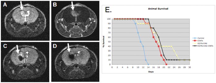
A., T2 weighted MRI showing a tumor xenograft with bright signal 7 days post tumor implantation (arrow); B., Tumor shown (arrow) by contrast enhancement after injection of the gadolinium contrast agent (Gd-DTPA); C., MRI signal drop (arrow) after CED of EGFRvIIIAb-IONPs; D., EGFRvIIIAb-IONP dispersion and T2 signal drop (arrow) on MRI 4 days after CED. E., Kaplan-Meier survival curve comparison of athymic nude mice after intracranial implantation of human U87ΔEGFRvIII cells and treatment by MRI guided CED of HBSS (control), IONPs, EGFRvIIIAb, or EGFRvIIIAb-IONPs. Statistical significance, P< 0.001, was estimated by log-rank method of CED of EGFRvIIIAb-IONPs, IONPs, and EGFRvIIIAb compared to HBSS CED.