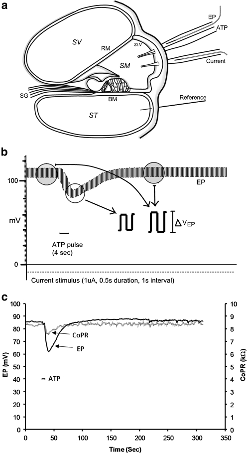
Fig. 1.
a Diagram showing the method for measuring the endocochlear potential (EP) and cochlear partition resistance (CoPR) from electrodes inserted through holes in the otic capsule into the scala media. b Trace of the EP showing a transient decline in EP with ATP microinjection (100 μM, 20 nL). Current pulses induced a change in voltage from which the CoPR was calculated. This voltage change was smaller during the introduction of ATP (reflecting decreased CoPR). c Changes in the CoPR and EP with time calculated for a single ATP microinjection (20 nL, 100 μM). SV scala vestibuli, ST scala tympani, SM scala media, RM Reissner’s membrane, St.V stria vascularis, BM basilar membrane, EP endocochlear potential SG spiral ganglion

