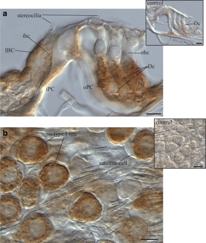
Fig. 3.
Detail of NTPDase5 immunolocalisation in adult rat cochlea. a NTPDase5-specific antibody showed strong supranuclear cytoplasmic immunoperoxidase labelling of Deiters’ cells (Dc). Weaker labelling was observed in the inner border cells (IBC), but the adjacent inner hair cell (ihc) was unlabelled. Residual labelling in head and footplate of outer pillar cells (oPC) was observed in control tissues after peptide block (inset). b Type I spiral ganglion neurons (based on their size and distribution), demonstrated strong NTPDase5-specific labelling in the cytoplasm, which was not observed in the surrounding satellite cells. Peptide block controls showed no immunolabelling in ganglion cell perikarya (inset). Scale bar 10 µm. Dc Deiters’ cell, iPC inner pillar cell, N nucleus, ohc outer hair cell, oPC outer pillar cell

