Fig. 3.
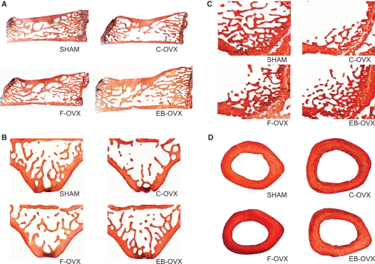
LM micrographs (50 μm thick) showing the bone histology from the four experimental animal groups after 30 days of treatment. (A) Sagittal sections of the 4th lumbar vertebra; (B) transversal sections of the 5th lumbar vertebra; (C) sagittal sections of the distal epiphysis of femur; (D) transversal sections at the mid-diaphyseal level of femur.

