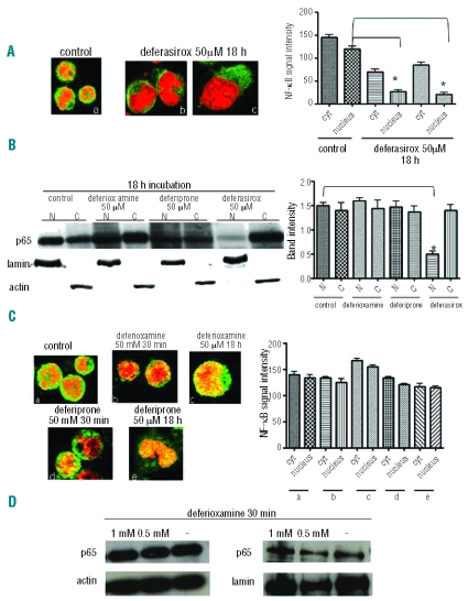
Figure 1.
Deferasirox inhibits NF-kB activity in K562 cells whereas deferioxamine and deferiprone do not. (A) Immunofluorescence assay using p65 antibody in control cells (a) and after incubation with deferasirox 50 mM for 18 h (b–c). Nuclei are stained in red, the green signal represents p65 subunit. In control cells the NF-κB subunit is mainly localized in the nucleus as indicated by the yellow signal whereas, after incubation with the drug, p65 subunit is localized in the cytoplasm in the inactive form as indicated by the green signal in the cytoplasm and the absence of yellow signal in the nucleus. The graph represents the signal intensity quantification in both the cellular compartments. Nuclear signal intensity is significantly different in control and incubated cells (P<0.001). (B) Western blotting using p65 antibody for the detection of proteins in either nuclear (N) or cytoplasmic (C) extracts in K562 cells. The upper line indicates the p65 antibody, while both the lower lines represent the internal controls for either cytoplasmic (actin antibody) or nuclear extracts (lamin antibody). p65 nuclear localization is decreased only after deferasirox incubation but neither deferioxamine nor deferiprone incubation. The graph shows the quantification of the signal intensity detected by Western blotting. (C) Immunofluorescence assay using p65 antibody in K562 cells before and after incubation with deferioxamine and deferiprone either at 0.5 mM for 30 min or 50 mM for 18 h. After all the different conditions of incubation, p65 remains localized in the nucleus in the active form as in control cells. The graph illustrates NF-κB signal intensity in all the different conditions of incubation. No statistically significant differences have been found between control values and K562 treated cells with both the drugs. (D) Western blotting using p65 antibody for the detection of proteins in either cytoplasmic or nuclear extracts in K562 cells. The upper line indicates the p65 antibody, while the lower line represents an internal control for either cytoplasmic (actin antibody) or nuclear extracts (lamin antibody). p65 nuclear localization is not decreased after deferioxamine incubation for 30 min.

