
Figure 1.
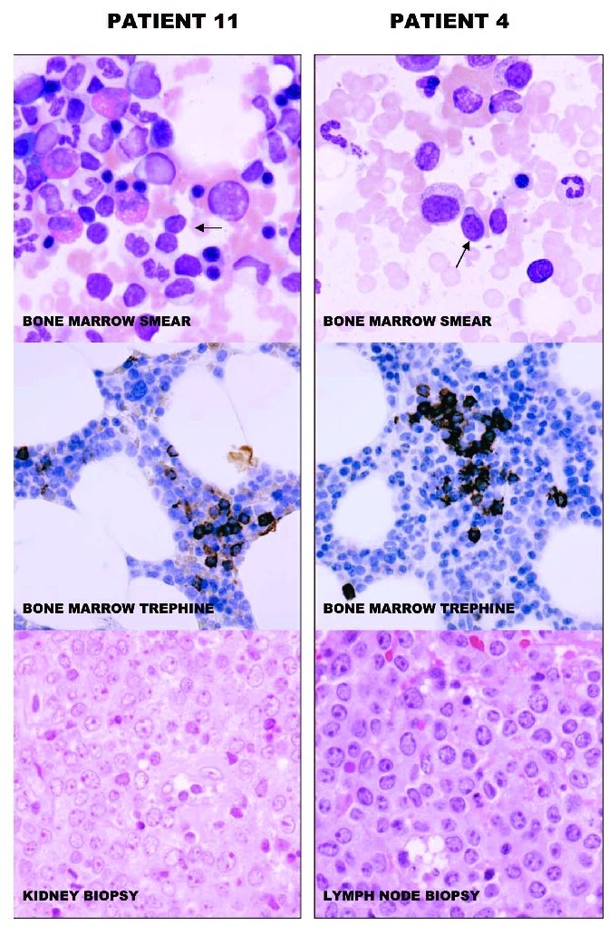
Wright’s-stained bone marrow smears (original x 600) and anti-CD20 stained sections of bone marrow trephines (original x 200) and hematoxylin and eosin stained tissue biopsies (original x 400) of patients 11 and 4 are illustrated. Small lymphoid cells (arrows), likely corresponding to MSBC are seen in the smears. The cells have round nuclei without prominent nucleoli and have varying amounts of cytoplasm. The cells of patient 4, having MSBC with a non-CLL-like phenotype have more cytoplasm than cells of patient 11, having MSBC with a CLL-like phenotype. The bone marrow trephines of both cases show sparse and small collections of small CD20+ B cells. The tissue biopsies show diffuse infiltration with large lymphoid cells with multiple nucleoli, typical of DLBCL. The images were acquired with an Olympus BX50 microscope (Olympus Corporation, Tokyo, Japan) equipped with a Hamamatsu C4742-95-10SC camera (Hamamatsu Photonics K.K., Hamamatsu City, Japan). The acquisition software was Paint Shop Pro 7.02 (Corel Corporation, Ottawa, Canada).