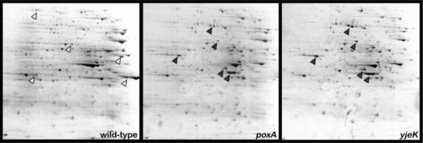
Figure 5. 2D SDS-PAGE of Salmonella poxA and yjeK mutants reveals similar protein expression profiles that differ from wild-type Salmonella.
Colloidal Coomassie-stained polyacrylamide gels after 2D gel electrophoresis on a PAGE gradient 8–16% acrylamide gel are shown. Isoelectric focusing was performed on a non-linear pH gradient ranging from 3–11. Arrows show prominent protein spots that are differ in expression between the wild-type strain (white arrows) and the yjeK/poxA mutants (grey arrows). Data on proteins displaying expression differences between the poxA mutant and wild-type are provided in Table 2 and Figure S5.

