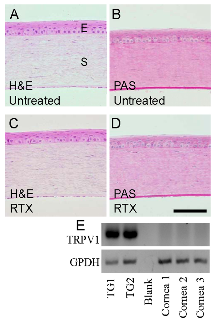
Figure 2.

RTX-treated and untreated corneas were histologically indistinguishable using H&E and PAS staining. Corneas were acquired 24 hr after treatment with 2 µg RTX, post-fixed in 10% formalin, and embedded in methyl methacrylate. Sections were stained with H&E or PAS and evaluated by a masked observer. “E” indicates the epithelial layer and “S” indicates the stromal layer. (A) H&E untreated; (B) PAS untreated; (C) H&E 2 µg RTX; (D) PAS 2 µg RTX. Scale bar = 200 µm. (E) RT-PCR shows TRPV1 transcripts are expressed in trigeminal ganglia (TG) but not the cornea, using GPDH as a normalizing control. Thus, RTX likely has no direct effect on resident corneal cells.
