
Figure 1.
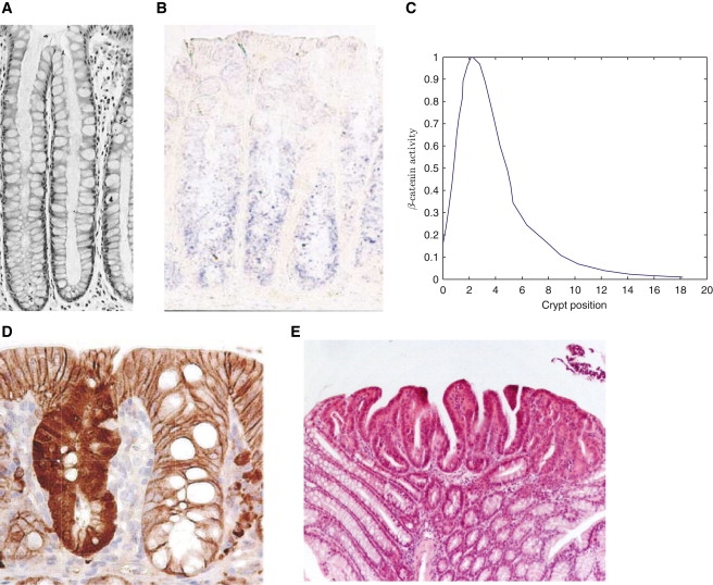
Experimental data on the crypt. (A) Section through a normal healthy crypt (12). (B) Wnt-6 stained murine colonic crypt (38). (C) Plot of normalized β-catenin activity along the crypt axis from a cross section through a mouse small intestinal crypt immunohistochemically stained for β-catenin expression. Data reproduced from Marshman et al. (39). Note that the presence of Paneth cells at the bottom of small intestinal crypts introduces features in the β-catenin profile that one may not expect to observe in colonic crypts. (D) A comparison of β-catenin distributions (stained) between an APC-mutated (left) and a normal (right) colonic crypt (taken from Barker et al. (40)). (E) Repopulation of neighboring crypts as a result of genetic mutations (13).