
Figure 6.
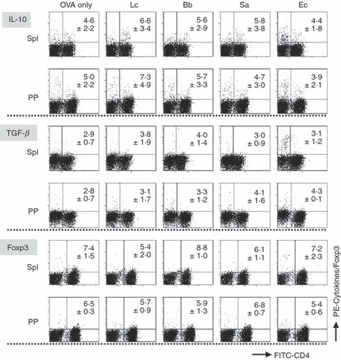
An analysis of the development of regulatory T cells from spleen and Peyer’s patch (PP) cells in response to various bacteria. Spleen and PP cells prepared from ovalbumin-specific T-cell receptor transgenic (OVA-TCR-Tg) mice were primarily cultured with or without (OVA only) various bacteria (10 μg/ml) in the presence of OVA for 6 days in order to induce the development of regulatory T cells. The cultured cells were harvested, re-stimulated with phorbol 12-myristate 13-acetate (PMA) plus ionomycin in the presence of brefeldin A for 4 hr and stained with fluorescein isothiocyanate (FITC)-labelled anti-CD4 and phycoerythrin (PE)-labelled anti-IL-10 or biotinylated anti-transforming growth factor (TGF)-β1 antibodies. Biotinylated antibody was detected using PE-labelled streptavidin. For the analysis of Foxp3-positive cells, the cells after the primary culture were stained with FITC-labelled anti-CD4 and PE-labelled anti-Foxp3 antibodies without re-stimulation. The stained cells were analysed by flow cytometry. The values represent the mean ± standard deviation of the percentages of cytokine-producing or Foxp3-expressing cells in the CD4+ T cells in three independent experiments. Lc, Lactobacillus casei; Bb, Bifidobacterium bifidum; Sa, Staphylococcus aureus; Ec, Escherichia coli.