
Figure 3.
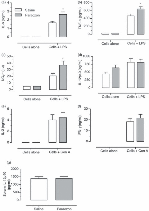
Functional responsiveness of lymphoid and myeloid cells. At the end of a 3-week exposure to saline or paraoxon, single spleen cell suspensions were cultured in the presence or absence of lipopolysaccharide (LPS; a–d), or concanavalin A (Con A; e,f). Culture supernatants were collected and tested for interleukin-6 (IL-6) (a), tumour necrosis factor-α (TNF-α) (b), nitric oxide (c), IL-12/IL-23p40 (d), IL-2 (e) or interferon-γ (IFN-γ) (f). Blood was also collected from each mouse and the serum level of IL-12/IL-23p40 protein was determined (g). Each data point represents the mean ± SEM of nine individually assayed mice per group. Asterisks denote significant differences between control and paraoxon groups (*P < 0·05). The results represent pooled data from three independent experiments.