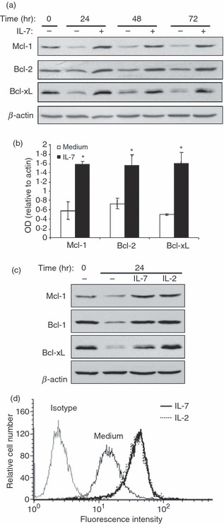
Figure 3.

Interleukin-7 (IL-7) up-regulates Bcl-2 proteins in human effector/memory T cells. (a) Cells at day 8 (0 hr time-point) were cultured under deprived conditions in the presence or absence of IL-7 (2 ng/ml) for the indicated periods of time. Whole cell lysates were then prepared and subjected to immunoblot analysis with specific antibodies against Mcl-1, Bcl-2, or Bcl-xL. The blot was stripped and re-probed with anti-β-actin monoclonal antibody (mAb) to ensure equal loading. The results are representative of five independent experiments performed with T cells from different blood donors. (b) Densitometric quantification of relative increase in Bcl-2 proteins in cells cultured for 24 hr under deprived conditions in the absence (medium) or the presence of IL-7. The results represent mean values (± SE) from three independent experiments and are expressed as the ratio between Mcl-1, Bcl-2 or Bcl-xL values and β-actin values. *P < 0·05 between IL-7-treated samples and non-treated samples (medium). (c) The cells were cultured under deprived conditions in the presence or absence of IL-7 (2 ng/ml) or IL-2 (50 U/ml) for 24 hr. The expression of Mcl-1, Bcl-2 and Bcl-xL was determined by immunoblot analysis. The results are representative of three independent experiments performed with T cells from different blood donors. (d) The cells were left unstimulated (medium) or stimulated with IL-7, IL-2 for 24 hr. The cells were then washed, stained with phycoerythrin-conjugated anti-Bcl-2 or phycoerythrin-conjugated isotypic control antibodies and analysed by fluorescence-activated cell sorting analysis. The control isotypic staining is shown for unstimulated cells, which is similar to control isotypic staining of IL-2- and IL-7-stimulated cells (data not shown). The results are representative of three different experiments performed with T cells from different blood donors.
