
Fig. 2. Neuroblasts do not express the D3R receptor in postnatal and adult brains.
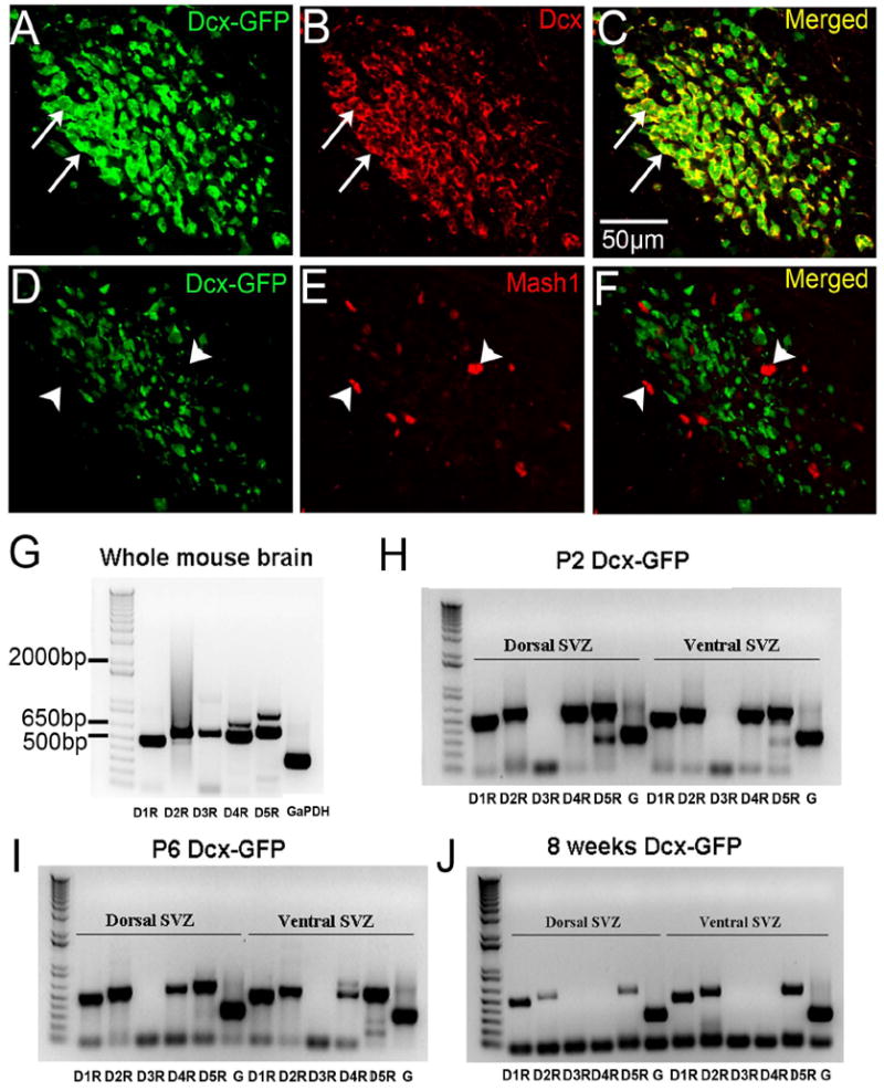
A-C: Dcx immunohistochemistry (red) in Dcx-GFP mouse coronal RMS sections. White arrows indicate examples of colocalized cells, note near perfect colocalization.
D-F: Mash1 immunohistochemistry (red) in Dcx-GFP coronal RMS sections. White arrow heads indicated examples of Mash1+ cells that are Dcx-GFP negative, note lack of colocalization.
G: Dopamine receptor RT-PCR from whole brains of Dcx-GFP mice. All five receptors were detected.
H-J: Dopamine receptor RT-PCR from GFP+ neuroblasts of P2 (H), P6 (I), and adult (J) Dcx-GFP mice. Note the D3R expression was not found in neuroblasts in any of the samples. G = GaPDH.