Figure 5.
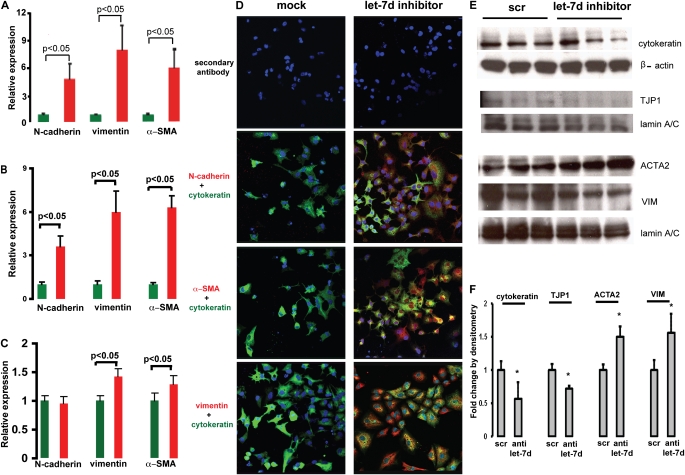
Inhibition of let-7d results in epithelial–mesenchymal transition changes. N-cadherin (CDH2), vimentin (VIM), and α-smooth muscle actin (ACTA2; α-SMA) mRNA levels were determined by quantitative real-time polymerase chain reaction in (A) A549 cells and (B) RLE-6TN cells 48 hours after transfection and in (C) NHBE cells 24 hours after transfection with 50 nM let-7d inhibitor. (D) Immunofluorescence imaging of A549 cells transfected with 50 nM let-7d inhibitor. Green fluorescence represents cytokeratin, an epithelial marker. Red fluorescence denotes the mesenchymal markers (CDH2, VIM, and ACTA2 [α-SMA]). Nuclei were counterstained with 4′,6-diamidino-2-phenylindole. Whereas red staining was observed in cells transfected with let-7d inhibitor (right), there was no staining in cells transfected with a control oligonucleotide (left). (E) Western blots of RLE-6TN cells transfected with 50 nM let-7d inhibitor. scr = scrambled sequence; TJP1 = tight junction protein-1. (F) Densitometric analysis of the immunoblots in (E), *P < 0.05. Fold change by densitometry is equivalent to the density of the targeted protein divided by the density of the corresponding housekeeping gene. Results represent averages and SD of triplicate experiments.

