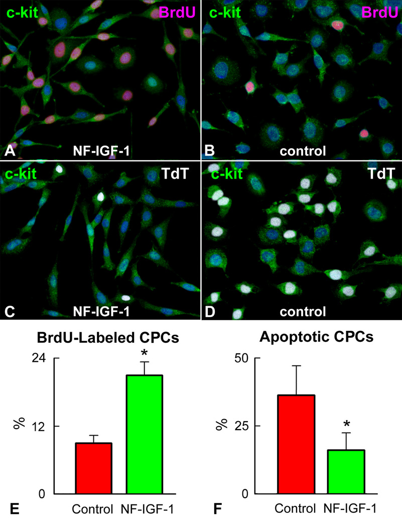
Figure 1.
IGF-1 and CPC growth and survival. CPCs (green) labeled by BrdU (magenta) are more frequent with (A) than without (B) NF-IGF-1. Conversely, TdT-labeling (white) is lower in CPCs exposed to NF-IGF-1 (C and D). CPCs incorporating BrdU (E) and undergoing apoptosis (F) in SFM and with NF-IGF-1. *Indicates P<0.05.

