
Figure 5. Colocalization of GAD 67 and 5-HT2CR in layer IV of the PrL.
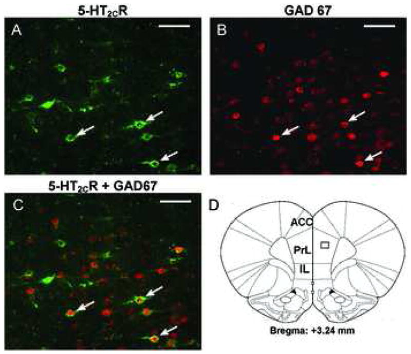
Photomicrographs of double-label immunofluorescent staining for the 5-HT2CR (green, A), and GAD 67 (red, B). Figure C displays the overlay of images in A and B to demonstrate colocalization. Approximately 50% of 5-HT2CR-positive cells in the field also contain GAD 67-IR. Examples of cells that express immunoreactivity for GAD 67 and 5-HT2CR are shown by arrows. D. Schematic drawing adapted from Paxinos and Watson (2004) corresponding to coronal sections (bregma 3.24 mm) from which images in A–C were captured. Scale bar: 50 μm. ACC: anterior cingulate cortex; PrL: prelimbic cortex; IL: infralimbic cortex.