
Figure 3.
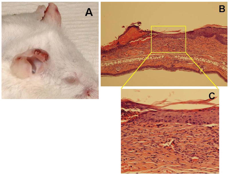
Development of cutaneous lesions in AE°DQ2 mice. (A) Erosions, ulcerations and crusting are present on the ears and muzzle. (B) H&E staining (10X) shows, thickening of the epidermis, hyperkeratosis as well as subepidermal blistering with hydropic degeneration and lymphoid infiltration in the subepidermal/dermal area (Reflective of 10 cutaneously affected mice). (C) Higher magnification (40x) shows the presence of vacuoles.