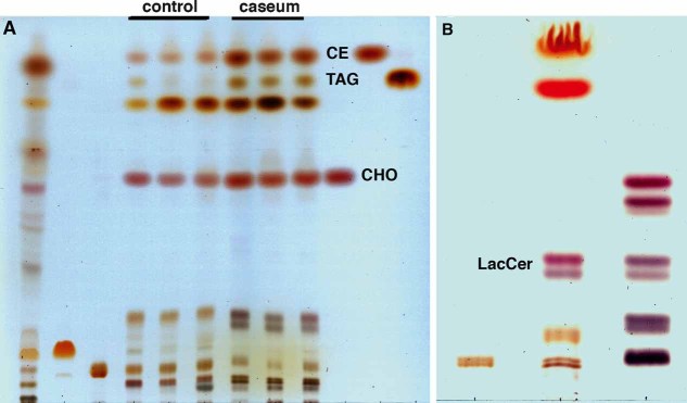
Figure 6. Thin-layer chromatographic analysis of lipids from the caseum and normal lung tissues.
- Dual solvent separation of granuloma lipids by TLC. Total lipids were extracted from normal lung tissues (lane 4–6, 120 µg) and caseous TB lung granulomas (lane 7–9, 120 µg), and then equivalent amounts were run in parallel with total lipids from Mtb CDC1551 (lane 1, 400 µg), and standard lipids, cardiolipin (lane 2, 50 µg), phosphatidylcholine (lane 3, 50 µg), CHOL (lane 10, 20 µg), CE (lane 11, 25 µg), TAG (lane 12, 20 µg). The lipids from the TB caseum exhibit an abundance of CHOL, CE, and TAG.
- Single solvent separation of lipid species with similar migration to the sphingolipids, illustrating the enrichment of LacCer in the caseum extract. Total lipids from the caseum (lane 2, 120 µg) were run together with sphingomyelin standard (lane 1, 5 µg) and neutral glycosphingolipids standard (lane 3, 30 µg). Caseous and fibrocaseous granuloma samples were derived from seven independent tissue samples, and normal lung tissue samples.

