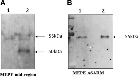
Figure 7.
Western blot analysis of primary dentin extracts with MEPE antibodies after 8% SDS-PAGE. A: MEPE mid-region. B: MEPE C-terminal (ASARM) region. Lane 1, control dentin; lane 2, vital hypophosphatemic dentin (patient 1). MEPE appears as a band at 55 kDa in a control dentin extract with both antibodies (A and B). In a hypophosphatemic sample, two clear bands are detected with the MEPE mid-region antibody at 55 and approximately 50 kDa (A), whereas a single 55-kDa band is detected with the ASARM antibody (B).

