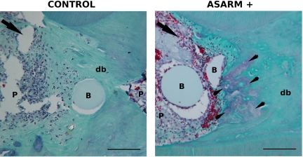
Figure 8.
P-ASARM peptide disturbs pulp repair in an injured rat pulp model. Beads soaked with the peptide (right panel) or with buffer (left panel) were implanted in the injured pulp of young rats. Animals were sacrificed at day 30 and demineralized maxillary blocks were observed after Masson’s trichrome staining. The arrowheads indicate purple-stained fibrous areas located in the green-stained reparative dentin bridge in ASARM animals. The arrow indicates the side of the pulp exposure. In the right panel, many red-stained erythrocytes are observed neighboring the beads. db, dentin bridge; B, bead; P, pulp. Scale bars = 100 μm.

