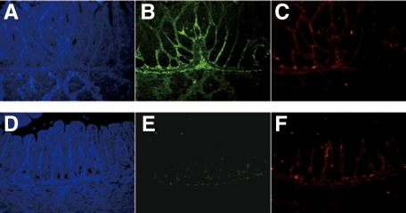
Figure 5.
Pyloric samples of normal mice (A, B, and C) and their slw/slw littermates (D, E, and F) were stained with PDE5A (green) and S-100 (red) for immunofluorescence study (×400 magnification). Note the inadequate PDE5A staining of the pylorus of the slw/slw mice compared with that of the normal mice. The blue staining was achieved with DAPI.

