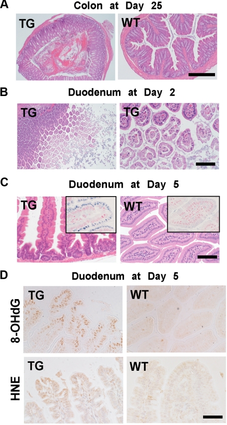
Figure 5.
Pathology of the intestine in SLC11A2 transgenic mice under an iron-rich diet. A: Low-power view of colon at day 25 after the start of an iron-rich diet. TGs revealed hemorrhagic erosion, whereas WT mice showed normal histology (Scale bar = 500 μm). B: Duodenum at day two of TG after the start of an iron-rich diet. Erosion of the apical surface of the duodenal epithelial cells was prominent (Scale bar = 400 μm and 100 μm, respectively). C: Iron deposits in the duodenal cells of survived mice at day five. Degenerative duodenal cells in TG accumulate iron in the cytoplasm, whereas those in wild-type do not (Scale bar = 140 μm). Inset, Perls’ iron staining (Scale bar = 70 μm). D: Oxidative stress in the duodenal cells at day five. Increase in nuclear 8-hydroxy-2′-deoxyguanosine (8-OHdG) and cytoplasmic 4-hydroxy-2-nonenal (HNE)-modified proteins were increased, indicating the pathological process is oxidative stress-dependent at day five (Scale bar = 70 μm).

