Figure 1.
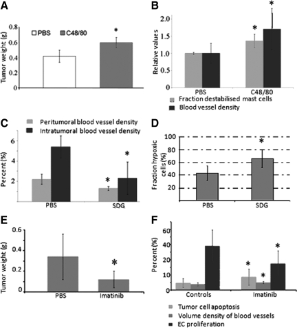
A: Coinjection of AT-1 cells with compound 48/80 stimulated tumor growth compared with PBS. *P < 0.05. B: Compound 48/80 stimulated mast cell degranulation (determined on toluidine blue staining and defined as mast cells with a discontinuous membrane) and increased the volume density of blood vessels. Values in A and B, are shown as means ± SD with six to ten animals in each group. *P < 0.01. C: SDG reduced the volume density of blood vessels both in the nonmalignant stroma surrounding the tumor and in the tumor stroma. *P < 0.05. D: SDG induced tumor hypoxia compared with PBS. The fraction hypoxic cells increased from 43 ± 11% to 66 ± 14%. *P < 0.05. Data from SDG experiment represent one experiment with three to seven animals in each group. E: Imatinib reduces growth of AT-1 tumors compared with controls. *P < 0.05. F: Significant effects on tumor cell apoptosis, volume density of blood vessels, and endothelial proliferation were observed after treatment with Imatinib. Values from experiment using Imatinib are shown as means ± SD with eight to ten animals in each group. *P < 0.05.

