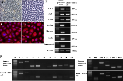
Figure 1.
Phase-contrast images (A and B) and immunocytochemical analysis for CK7 (C) and CK19 (D) in hTERT-HPDE cells cultured with serum-free conditioned medium. hTERT-HPDE cells show a small cobblestone appearance and are stained with CK7 and CK19. Scale bars: 80 μm (A); 40 μm (B); 20 μm (C and D). E: RT-PCR for CA-II, CK7, CK19, amylase, glucagon, insulin, and somatostatin in hTERT-HPDE cells. hTERT-HPDE cells express mRNAs of CA-II, CK7, and CK19. Human pancreatic tissues were used as positive controls. F: RT-PCR for tight junction molecules in hTERT-HPDE cells. mRNAs of claudin-1, -4, -7, and -18, occludin, JAM-A, ZO-1, ZO-2, and tricellulin are detected in hTERT-HPDE cells. Human pancreatic tissues were used as positive controls. CL, claudin; Oc, occludin; TRIC, tricellulin; M, 100-bp ladder DNA marker.

