Figure 1.
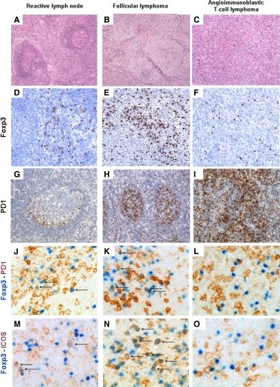
Immunohistochemistry in lymph node biopsies. Reactive lymph node: A (×100) and D (×200): FOXP3+ cells are scattered within the interfollicular zones. G (×100): PD1+ cells are mostly located in the germinal center. J and M (×400): Double staining with FOXP3 (nuclear) and PD1 (membrane) or FOXP3 and ICOS (membrane) show few cells expressing both markers (arrows). Follicular lymphoma: B (×100) and E (×200): Numerous FOXP3+ cells surround the nodules. H (×100): Many cells with the follicles are PD1+. K and N (×400): A significant proportion of cells are double stained for FOXP3 and PD1 or ICOS (arrows). AITL: C (×100) and F (×200): FOXP3+ cells are rare and scattered. I (×100): Many cells are PD1+, including AITL neoplastic cells. L and O (×400): Double staining with FOXP3 and PD1 or FOXP3 and ICOS do not show double-positive cells.

