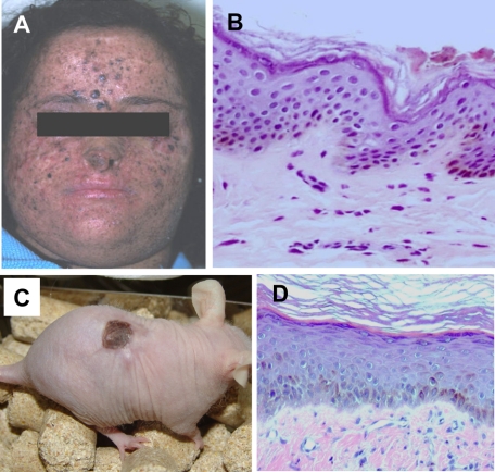
Figure 6.
Development of a XP-C skin-humanized mouse model. A: Physical characteristic appearance of one of the XP-C donor patients. B: Histological appearance (H&E staining) of a section from the patient’s skin biopsy used to culture skin cells. C: Appearance of a representative XP-C regenerated skin-engrafted mouse. Note that cells from patient 1 (shown in A) give rise to pigmented regenerated skin. D: Histological appearance (H&E staining) of a section of the regenerated human skin in a mouse engrafted with cells from patient in A.

