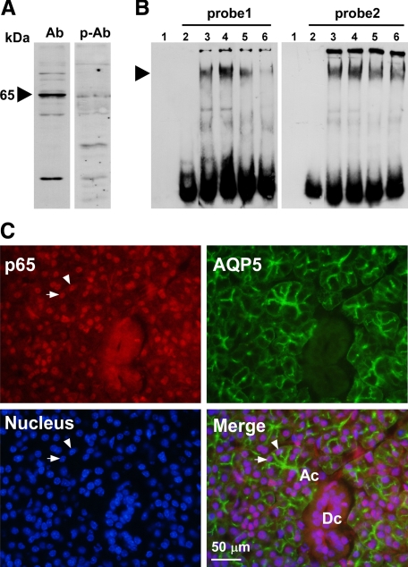
Figure 6.
Analysis of NF-κB in the PG. A: Western blotting of the NF-κB protein in a PG nuclear extract from a C3H/HeN mouse injected with LPS 1 hour before sacrifice. Ab indicates anti-p65 polyclonal antibody; p-Ab, peptide-preabsorbed anti-p65 polyclonal antibody. B: EMSA of NF-κB in the PG from nontreated and LPS-injected C3H/HeN mice. probe1, 5′-AGTCTCAGGCACTTCCCTAAGCC-3′; probe 2, 5′-ACTCCCGATCCACTCCCCCGCTCC-3′. Lane 1, no probe and no protein sample; lanes 2–6, experiments in the presence of probe; lane 2, no protein sample; lane 3, nuclear extract from nontreated C3H/HeN mice; lane 4, nuclear extract from LPS-injected C3H/HeN mice; lane 5, nuclear extract from the LPS-injected C3H/HeN mice preincubated with 50-fold molar excess of unlabeled double-stranded oligonucleotide of NF-κB specific probe; lane 6, nuclear extract from an LPS-injected C3H/HeN mouse, with extract preincubated with antibody against p65. An arrowhead indicates the shifted double strand DNA probes. C: Immunohistochemical detection of p65 and AQP5 in the PG of a C3H/HeN mouse that had been injected with LPS 1 hour before sacrifice. Subcellular localization of p65 protein by Alexa Fluor 594 (red, upper left), subcellular localization of AQP5 by FITC immunofluorescence (green, upper right), cell nuclei stained by DAPI (blue, lower left), and a merged picture (lower right) are shown. Ac indicates an acinus; Dc, a duct. Arrowheads indicate typical nuclei of acinar cells showing exact localization of p65 in the nucleus.

