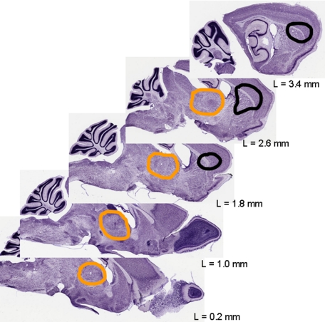
Figure 1.
Simulation of the localization and extent of lesions after the excitotoxic insult. The primary excitotoxic lesion includes the striatum and fronto-lateral parts of the cortex (borders marked in black). The zone of secondary neurodegeneration in the thalamus is encircled in orange. Adapted to Nissl-stained sections from BrainMaps.org. L indicates laterality.

