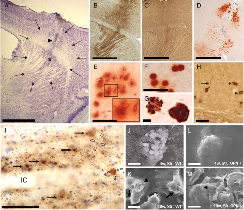
Figure 2.
Visual aspects of the corticostriatal lesion. A: The arrows indicate the boundaries of a typical corticostriatal neuron-depleted area in a Cresyl-stained section. The arrowhead indicates the needle tract, the asterisk a small region of central necrosis demarcating the position of the needle-tip. Microglial (B) and astroglial reaction (C) were visualized in iba1- and GFAP-stained sections, and microcalcification with Alizarin Red S stain (D). E: Type 1 (nascent) deposits are often neuron-shaped and have prominent halos (see also inset). F: Type 3 (mature) deposits are round, dense, and have almost no halo. G: Double staining with Alizarin Red S (for calcium deposits) and LF123 (for OPN) indicates a dense association of OPN with calcified material. Note the “bulby” surface of the left deposit. H: Osteopontin (OPN)-stained structures without (arrowheads) and with processes reminding of neurons (arrow) represent osteopontin-positive calcium deposits. I: Section counterstained with Cresyl violet depicts presence of OPN in some neurons (long arrows) where it fills rather homogenously the cytoplasm. Most OPN, however, is found in small-dotted fashion in the neuropil but not in the white matter of the internal capsule (IC). Occasionally dots mark the outer surface of large perikarya (open pointers). An OPN-stained extracellular deposit is associated with small cresyl-positive structures representing most probably microglial nuclei (open arrows). J: In SEM, mainly flat and angular bulbs are observed at the surface of a corticostriatal deposit of a wild-type mouse four weeks after the excitotoxic lesion. K: After ten weeks, these bulbs are often interconnected with bridges (arrows), and parts of the surface of the bulbs appear rough (arrowhead). In OPN knockout mice, corticostriatal deposits have flat and smooth surfaces (arrowhead) without obvious difference between four (L) and ten weeks survival time (M). OPN−/− indicates osteopontin-deficient mouse; w, weeks; WT, wild-type mouse. Scale bars: 1 mm (A–D); 50 μm (E, F, H, I); 5 μm (G, J, K, L, M).

