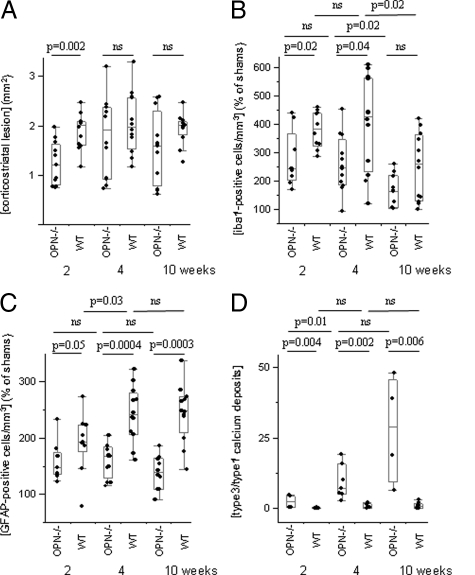
Figure 3.
Morphological and morphometric analyses of the corticostriatal lesion. A: The corticostriatal lesion was larger in wild-type than in OPN-KO mice two weeks after the excitotoxic insult but did not differ significantly at later time points. B: Number of microglial cells was higher in wild-type than in OPN-KO mice two and four weeks after lesioning, and returned to lower levels after ten weeks, now without significant difference between the strains. C: GFAP-positive cells were more numerous in wild-type than in OPN-KO mice at all time points investigated. D: The ratio of the deposits (type3/type 1) was higher in OPN-KO than in wild-type mice at all investigated time points.

