Figure 5.
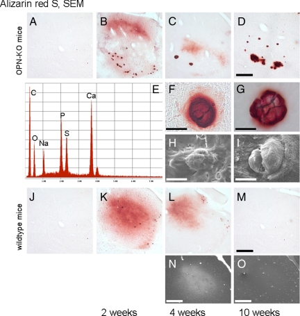
Characteristics of thalamic microcalcification. Alizarin Red S: (A, J) Sham animals with survival times of two weeks, without thalamic microcalcification (note that a considerable percentage of OPN-KO sham animals developed detectable thalamic microcalcification in particular after 10 weeks survival, see text). Thalamic microcalcification occurs two and four weeks after corticostriatal injury as Alizarin Red S-positive misty staining, without major differences between the strains (OPN-KO mice: B, C; wild-type mice: K, L). The misty staining disappears after four weeks (D, M). Dense deposits are almost only found in OPN-KO mice (D). They are observable two weeks after the excitotoxic corticostriatal lesion and present as even larger aggregates after four and ten week survival time (B–D). A dense aggregate with a small halo from an OPN-KO mouse with four weeks survival is shown with higher magnification (F). Halos have disappeared ten weeks after the corticostriatal excitotoxic lesion (G). SEM: (H, I) Two representative thalamic deposits of OPN-KO mice are shown: the surface appears flat and smooth, without obvious differences between four and ten weeks survival time. Energy dispersive (EDS) X-ray analysis in the SEM of a thalamic deposit shows high Ca- and P-peaks (E; similar results were obtained for corticostriatal deposits of both wild-type and OPN-KO mice, not shown). The positive signal in wild-type mice with four weeks survival in the back-scatter mode (N) is no longer found ten weeks after the corticostriatal lesion, and larger deposits are absent (O). SEM indicates scanning electron microscopy. Scale bar = 300 μm, except single deposits of the OPN-KO mice = 10 μm.

